
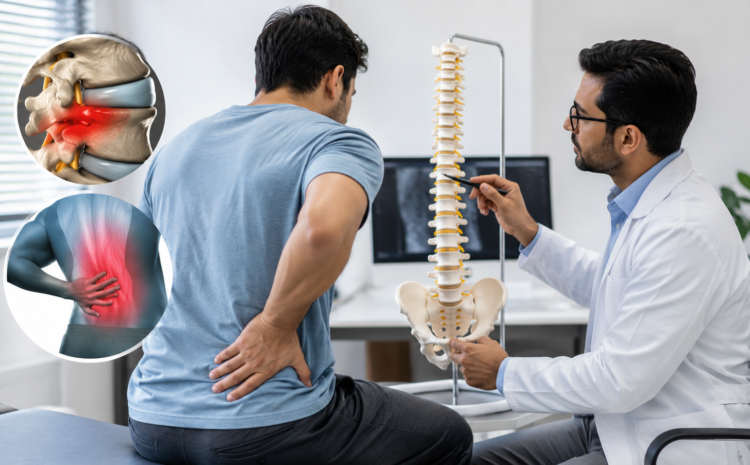
You woke up with severe back pain, searched your symptoms online, and now you’re somehow more confused than when you started. Sound familiar? Back pain is one of the most common reasons people visit a doctor, and two of its most frequently confused causes are a slipped disc and a muscle spasm. On the surface they can feel remarkably similar — both can stop you in your tracks, make it impossible to get comfortable, and leave you wondering if something is seriously wrong. But underneath, they are entirely different conditions with different causes, different diagnostic paths, and most importantly, different treatments. Getting the right diagnosis is not a formality — it is the difference between recovering quickly and making things significantly worse. This blog explains exactly how doctors tell them apart.
Understanding the Basics: What Is a Slipped Disc?
Your spine is made up of bones called vertebrae stacked on top of each other, and between each pair of vertebrae sits a disc — a tough outer ring (the annulus fibrosus) surrounding a soft, gel-like center (the nucleus pulposus) that acts as a shock absorber and allows your spine to flex and bend. A “slipped disc” is a colloquial term for what doctors call a disc herniation — when the soft center pushes through a tear or weakness in the outer ring.
Here is what you need to understand about disc herniations:
- The disc does not literally “slip” out of place — rather, the inner material protrudes through the outer casing in varying degrees, from a mild bulge to a full extrusion where disc material breaks free into the spinal canal.
- The lumbar spine (lower back) is the most commonly affected region, particularly the L4-L5 and L5-S1 levels — the two lowest discs bear the most load and are subject to the greatest mechanical stress; the cervical spine (neck) at C5-C6 is the second most common site.
- A bulging disc means the outer wall is intact but distorted, like a burger patty pressed slightly beyond its bun; a herniated disc means the outer wall has torn and inner material is pushing through — the latter tends to cause more severe and radicular (nerve-related) symptoms.
- The disc itself has limited pain sensation, but when it presses on a nearby nerve root — the point where a spinal nerve exits the vertebral column — it triggers a cascade of pain, numbness, tingling, and weakness that can travel the entire length of that nerve into the leg or arm.
- Disc herniations are most often caused by gradual degeneration over time combined with a triggering event such as a lifting injury, though they can also occur suddenly in younger people during high-load activities.
Understanding the Basics: What Is a Muscle Spasm?
A muscle spasm is an involuntary, sustained contraction of one or more muscles that does not release on its own. In the back, this most often affects the large paraspinal muscles — the thick bands of muscle running on either side of your spine — and can produce pain that feels alarmingly severe given that no structural damage has occurred.
Here is what drives muscle spasms and why they are so often misunderstood:
- Common triggers include a sudden awkward movement, prolonged poor posture, muscle fatigue, physical overexertion, dehydration (muscles require adequate fluid and electrolytes to function), and even emotional stress, which increases baseline muscle tension throughout the body.
- The pain of a muscle spasm can be genuinely intense — disproportionately so relative to the underlying cause — because the contracted muscle compresses its own blood supply, causing a buildup of metabolic waste products that stimulate pain receptors in a reinforcing cycle.
- Acute muscle spasms come on suddenly, often in response to a specific movement, and resolve within days to weeks; chronic muscle tension develops gradually from sustained poor posture or repetitive strain and tends to be duller but more persistent.
- Muscle spasms are far more common than disc herniations — the majority of acute back pain episodes seen in clinical practice have a muscular rather than structural origin, which is why assuming a disc problem without a proper examination often leads patients down the wrong treatment path.
- Without nerve involvement, muscle spasm back pain stays local — it does not travel down the leg, does not cause numbness, and does not produce the electrical, burning quality that nerve compression does.
Symptom Comparison: How the Pain Feels Differently
This is often where patients begin to recognize their own experience. Read both lists carefully, because the differences are genuinely distinct when you know what to look for.
Slipped Disc Symptoms:
- Radiating pain that travels down one leg or one arm — often described as shooting, burning, or electric — is the hallmark of nerve root compression from a herniated disc and is known as sciatica when it involves the sciatic nerve in the lower back.
- Numbness, tingling, or a pins-and-needles sensation in a specific part of the leg, foot, arm, or hand indicates that a nerve is being compressed, and the location of those sensations often tells doctors exactly which spinal level is involved.
- Weakness in a specific muscle group — difficulty lifting the foot, pushing off the toes, or gripping with the hand — suggests that nerve compression has progressed to the point of affecting motor function, which is a more urgent finding.
- Pain that worsens significantly with sitting, coughing, sneezing, or bending forward is characteristic of disc-related pain because these actions increase pressure within the disc and on the compressed nerve root.
- Bladder or bowel changes — including difficulty urinating, incontinence, or loss of sensation in the groin area — alongside back pain constitute a medical emergency requiring immediate attention, as they may indicate cauda equina syndrome (compression of the bundle of nerves at the base of the spine).
Muscle Spasm Symptoms:
- Sharp, cramping, or knotted pain localized to a specific area of the back — often describable with a single finger rather than a vague ache across the whole back — is typical of muscular involvement.
- A visible or palpable area of hardness or tightness in the muscle, sometimes a discrete tender knot called a trigger point, can often be felt by pressing on the affected area.
- Pain that worsens with movement but stays entirely within the back and does not travel to the legs, arms, or extremities in any form strongly suggests a muscular rather than neurological cause.
- Stiffness that eases with gentle heat, light stretching, or moving around after an initial period of rest is a typical pattern for muscle spasm — the contracted tissue responds to warmth and movement in ways that nerve compression does not.
- No neurological symptoms — no numbness, no tingling, no weakness in the limbs, and no changes to bladder or bowel function — is one of the most reassuring features suggesting a purely muscular problem.
How Doctors Diagnose the Difference: The Clinical Examination
A skilled spine specialist can differentiate between a muscle spasm and a disc herniation in most cases through a structured physical examination alone — often before any imaging is ordered. Understanding what your doctor is looking for makes the appointment more productive.
- The straight leg raise (SLR) test involves lying flat while the doctor slowly lifts your straightened leg; reproduction of the radiating leg pain (not just back tightness) before 60 degrees of elevation is a positive result that suggests nerve root compression from a herniated disc at the L4, L5, or S1 level.
- Deep tendon reflex testing — tapping the knee and ankle tendons to check reflex responses — identifies which nerve root is affected; a reduced or absent ankle jerk suggests S1 nerve involvement, while a diminished knee jerk points to L4.
- Dermatomal sensory testing maps sensation across specific regions of the skin, each corresponding to a particular spinal nerve level — an area of reduced sensation tells the doctor which disc is likely responsible for the compression.
- Muscle strength grading of specific movements (extending the big toe, pushing down through the foot, raising the heel) identifies whether nerve compression has progressed to motor weakness.
- Palpation of the paraspinal muscles — pressing firmly along either side of the spine — reveals the board-like hardness of a muscle in spasm versus the bony tenderness over a vertebra that might indicate a fracture or structural problem; muscles in spasm are palpably different from normal resting muscle tissue.
- The Spurling test is used when cervical disc involvement is suspected: the doctor gently presses down on the top of your head while it is tilted slightly to one side — reproduction of radiating arm pain is a positive result suggesting nerve root compression in the neck.
Diagnostic Tools: When Imaging Is Needed and When It Is Not
One of the most common sources of confusion for patients is why their doctor may not immediately order a scan. Understanding the reasoning makes this decision far less frustrating.
- Immediate imaging is not indicated for most acute back pain because the majority of episodes — including many disc herniations — resolve with conservative treatment within six weeks, and early imaging rarely changes initial management while exposing patients to unnecessary cost and radiation.
- X-rays are useful for identifying fractures, spondylolisthesis (a vertebra slipping forward on the one below it), significant bone spurs, and gross structural deformity, but they cannot visualize discs, nerves, or soft tissue.
- MRI (magnetic resonance imaging) is the gold standard for diagnosing disc herniation — it provides detailed images of the disc, the nerve roots, the spinal cord, and surrounding soft tissue without radiation, and is typically ordered when neurological symptoms are present or when symptoms persist beyond four to six weeks of conservative treatment.
- CT scanning provides excellent bony detail and is used as an alternative when MRI is contraindicated (for example, in patients with certain implants or severe claustrophobia), though it offers less soft-tissue detail and involves radiation.
- EMG (electromyography) and nerve conduction studies measure the electrical activity of nerves and muscles and can confirm both the presence and the extent of nerve damage from disc compression — particularly useful when the clinical picture and MRI findings don’t perfectly align.
- A critically important truth for patients: a significant proportion of people over 40 have disc bulges or herniations visible on MRI with absolutely no symptoms — this is why imaging findings must always be interpreted alongside the clinical examination, and why a scan finding alone does not define your diagnosis or determine your treatment.
Treatment Differences: Why Correct Diagnosis Changes Everything
This is where accurate diagnosis translates directly into outcomes. Treating a muscle spasm as though it were a disc herniation — or vice versa — either delays recovery or risks making the condition worse.
Treating a Muscle Spasm
- Short-term relative rest followed by early gentle movement is the recommended approach — complete bed rest for more than 48 hours has been shown to prolong spasm recovery rather than help it, as sustained immobility further stiffens the muscle.
- Heat therapy applied to the affected area for 15 to 20 minutes at a time relaxes contracted muscle fibers and increases local blood flow, accelerating the clearance of the metabolic waste products driving the pain cycle.
- Muscle relaxant medications (such as cyclobenzaprine or methocarbamol) are useful for acute severe spasms that are preventing sleep or normal movement, and are typically prescribed for short periods only.
- NSAIDs (non-steroidal anti-inflammatory drugs like ibuprofen or naproxen) reduce both pain and the inflammation that often accompanies sustained muscle contraction.
- Physiotherapy focusing on gentle stretching, progressive strengthening of the core and paraspinal muscles, and postural correction addresses the mechanical drivers of spasm and significantly reduces the risk of recurrence.
- Trigger point therapy and dry needling — techniques in which a therapist manually releases or needles a hyperirritable knot in the muscle — can produce rapid and significant relief for chronic or recurrent spasm patterns that do not respond to stretching alone.
Treating a Slipped Disc
- Conservative management is always the starting point for disc herniation treatment — a structured program of rest, anti-inflammatory medication, physiotherapy, and time resolves the majority of disc herniations without surgery, as the herniated material is gradually reabsorbed by the body.
- Specific physiotherapy protocols — including the McKenzie method (extension-based exercises that centralize radiating pain) and neural mobilization techniques (gentle movements that mobilize the nerve within its sheath) — are more effective for disc-related nerve pain than generic back exercises.
- Epidural steroid injections (injecting anti-inflammatory corticosteroid medication into the space around the spinal cord) can provide significant temporary relief from nerve root pain, allowing patients to engage more effectively with physiotherapy.
- Surgical options — including microdiscectomy (keyhole removal of the herniated disc material) and endoscopic discectomy — are considered when conservative treatment has not provided adequate relief after six to twelve weeks, or when neurological deficits are progressing.
- Emergency surgical referral is required immediately when a patient develops foot drop (inability to lift the front of the foot), progressive leg weakness, or any change in bladder or bowel function — these signs indicate serious nerve compromise that requires urgent decompression.
When Back Pain Is Neither — Other Conditions Doctors Rule Out
Not all back pain comes from discs or muscles, and part of good back pain diagnosis is systematically excluding other causes.
- Sacroiliac joint dysfunction — inflammation or instability of the joint connecting the spine to the pelvis — causes localized buttock and lower back pain that closely mimics both disc and spasm pain and requires specific provocation tests to identify.
- Piriformis syndrome occurs when the piriformis muscle in the buttock compresses the sciatic nerve, producing sciatica-like leg pain that can be indistinguishable from disc-related sciatica without careful examination.
- Facet joint arthritis causes a characteristic pattern of pain that is worse with extension (bending backward), eases with sitting and forward flexion, and is often most severe first thing in the morning.
- Vertebral compression fractures — particularly in older patients or those with osteoporosis — can cause sudden, severe back pain that is entirely mechanical in origin; a history of osteoporosis combined with sudden pain after minimal activity should prompt immediate imaging.
- Kidney stones or infections can refer intense pain to the back in a pattern that mimics muscular pain, which is why the location, character, and associated symptoms (such as pain with urination or fever) are part of every back pain assessment.
Red Flags: When Back Pain Needs Emergency Attention
These symptoms should prompt you to seek same-day or emergency medical evaluation — do not wait for a scheduled appointment:
- Sudden loss of bladder or bowel control, or an inability to urinate, alongside back pain may indicate cauda equina syndrome — a surgical emergency with a narrow treatment window.
- Progressive weakness or numbness in both legs rather than one side, or a rapidly spreading neurological deficit, requires emergency spine imaging and assessment.
- Back pain following significant trauma — a road accident, a fall from height, or a sports collision — warrants urgent imaging to exclude fracture or spinal instability.
- Fever combined with back pain raises the possibility of spinal infection (discitis or epidural abscess), particularly in patients who have recently had spinal procedures, IV drug use, or immunosuppression.
- Unexplained weight loss alongside back pain is a red flag combination that must be investigated to exclude malignancy involving the spine.
- Pain that is constant, worsening, and completely unrelieved by any position — particularly at night — is atypical of both mechanical disc and muscle problems and requires investigation for non-mechanical causes.
Prevention: How to Protect Your Spine and Muscles Long-Term
Whether your back pain was muscular or disc-related, the strategies for preventing recurrence overlap substantially.
- Core strengthening — specifically targeting the deep stabilizing muscles (transversus abdominis, multifidus) rather than simply the visible surface muscles — is the single most evidence-supported intervention for preventing both disc injury and recurrent muscle spasm.
- Correct lifting technique means bending at the knees, keeping the load close to your body, and never twisting your spine while bearing weight — the combination of axial load and rotation is the most common mechanism for disc herniation.
- Ergonomic workstation setup with a properly adjusted chair height, monitor at eye level, and lumbar support prevents the sustained flexed posture that increases disc pressure and fatigues paraspinal muscles over hours of sitting.
- Movement breaks every 30 to 45 minutes during prolonged sitting reset disc pressure, restore muscle circulation, and interrupt the pattern of sustained posture that leads to both spasm and disc degeneration over time.
- Staying well-hydrated preserves the water content of your spinal discs — dehydrated discs lose height and shock-absorbing capacity, making them more vulnerable to herniation under load.
- Yoga and Pilates combine flexibility, core stability, and body awareness in ways that specifically support spinal health and have meaningful evidence for reducing back pain recurrence.
Conclusion
Back pain — whether it comes from a slipped disc or a muscle spasm — should never be left to self-diagnosis or treated based on assumption alone. Both conditions are highly treatable when identified correctly, and both can be made significantly worse by the wrong treatment. An accurate diagnosis from a qualified spine specialist is not just a formality — it is the most important step in your recovery. If you have been managing back pain on your own, or if new symptoms have appeared that worry you, do not wait for them to escalate. Book a spine consultation today and get a diagnosis that actually guides your treatment in the right direction.
Frequently Asked Questions
Q: Can a muscle spasm cause as much pain as a slipped disc?
Yes — and this surprises many people. A severe acute muscle spasm can be genuinely debilitating, producing pain intense enough to prevent standing, sleeping, or moving normally. The mechanism is different from disc pain (no nerve compression is involved), but the subjective pain experience can be equally severe. In some cases, patients with pure muscle spasms report pain they rate as 9 or 10 out of 10 on a pain scale. The distinction that matters clinically is not the intensity of the pain but its character — the presence or absence of neurological symptoms like radiating pain, numbness, or weakness distinguishes the two and determines the appropriate treatment path.
Q: How long does it take to recover from a slipped disc vs. a muscle spasm?
Recovery timelines differ significantly. An acute muscle spasm in an otherwise healthy person typically resolves within three to fourteen days with appropriate management — heat, gentle movement, anti-inflammatory medication, and physiotherapy. Most people are functionally back to normal within two to four weeks. Disc herniation recovery is more variable: around 90 percent of patients with a herniated disc improve with conservative treatment over six to twelve weeks, with the herniated material gradually reabsorbing over months. Neurological symptoms (numbness, tingling) may take longer to fully resolve than pain, sometimes persisting for several months even after the disc has healed. Cases requiring surgery followed by rehabilitation extend the timeline further.
Q: Can I exercise with a slipped disc or will it make things worse?
The answer depends entirely on which exercises and at what stage of recovery. Complete inactivity is not recommended for disc herniation — prolonged bed rest weakens core muscles and slows recovery. However, the wrong exercises can significantly worsen nerve compression: traditional sit-ups, heavy deadlifts, toe touches, and any exercise that loads the spine in flexion are contraindicated in the acute phase of lumbar disc herniation. The appropriate exercises — walking, McKenzie extension movements, specific neural mobilization techniques — must be guided by a physiotherapist who has assessed your specific disc level and neurological status. Never begin an exercise program for a slipped disc without professional guidance.
Q: Is an MRI always necessary to diagnose a slipped disc?
No — and in many cases, especially in the first four to six weeks of symptoms, MRI is not the first step. A skilled spine specialist can make a clinical diagnosis of probable disc herniation based on the pattern of your symptoms, the results of the neurological examination, and the straight leg raise test. MRI is typically ordered when: neurological symptoms (numbness, weakness) are present; symptoms persist beyond four to six weeks without improvement; the clinical picture is ambiguous; or surgery is being considered and the exact level of herniation needs to be confirmed. The important caveat is that MRI findings must always be interpreted alongside the clinical examination — an incidental disc bulge on MRI means very little without corresponding symptoms.
Q: Can a slipped disc heal on its own without surgery?
Yes — and for the vast majority of patients, it does. Research consistently shows that approximately 90 percent of people with disc herniation recover with conservative treatment alone over six to twelve weeks, without surgical intervention. The herniated disc material is gradually broken down and reabsorbed by the body’s immune cells, and the nerve root inflammation settles as the mechanical irritation reduces. The factors that most strongly predict the need for surgery are: progressive neurological deficits (increasing weakness), cauda equina syndrome (bladder/bowel involvement), or failure to improve after a genuine trial of six to twelve weeks of well-managed conservative treatment. Surgery, when it is indicated, is highly effective — but it is the exception, not the rule.